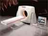
The big “oops” of the week in the medical community concerns CT scanning, otherwise known as computed tomography or CAT Scaning (computer aided tomography). Touted as miracle technology when it first hit the scene in the 1970s (in fact, the developers of modern CT scan technology shared the Nobel Prize in 1979), CT scans provide three-dimensional images far more detailed and flexible than normal X-rays. Doctors love to rely on them, and so the number of CT scans performed in the US increased 23-fold from three million in 1980 to about 70 million by 2007. At a typical cost of $500 and up to $1500 per scan, which can mean $6000 or more for diagnostic scans of several body parts, it’s been a profitable addition to the medical bag of tricks.
Despite assurances from doctors that the benefits of getting such accurate diagnostic images outweigh the risks, new evidence indicates that the radiation received from CT scans increases cancer risk…by a substantial margin. A study just completed at four San Francisco hospitals found the median dose of radiation delivered during CT scans was higher than previously thought. Also, the radiation levels varied wildly for the same procedure from hospital to hospital and even within the same institution. In fact, there was a 13-fold difference between the highest and lowest doses of radiation for identical procedures.
To put the radiation exposure in perspective, a complete set of dental x-rays (about 18 exposures) provides an effective dose of about 1.5 mrem (or .015 mSv). The average person absorbs one millisiervert (mSv) of background radiation annually just from routine living. A single pelvic/abdominal CT scan, on the other hand, exposes the patient to an average of 31 mSv while a typical head scan puts out an average of two mSv. But those are just averages. The researchers discovered that while a head scan at one hospital exposed patients to only 0.3 mSv, the same procedure at another hosptial exposed patients to six mSv (about 400 times the exposure of a complete set of dental x-rays). The deviations become startling when considering variations in the radiation for the abdominal/pelvic series, where researchers found radiation as high as 90 mSv at one hospital — almost 60 units above the average. This represents a 13-fold variation in dose and means that some patients are getting far more exposure than necessary for an adequate image. Consider that the survivors of Hiroshima suffered increased cancer risk from radiation exposures estimated to be as low as 10 mSv, and that the typical CT scan delivers as much radiation as 74 mammograms and 442 chest X-rays.
Given these figures, it’s no wonder that a second study published in the Archives of Internal Medicine concluded that 29,000 new cancers would result from CT scans performed in 2007 alone. The majority of those cancers (14,000), not surprisingly, would result from scans of the abdomen and pelvis, according to the projections. Another 4,100 would come from chest scans; 4,000 from head scans; and 2,700 from CT angiograms. The most vulnerable populations include younger adults and women. The researchers project that those aged 35-54 would develop one-third of the projected cancers while women would develop two-thirds of those incipient cancers.
Viewed through another lens, researchers estimate that one out of every 270 women and one of every 600 men who received a CT coronary angiogram at age 40 would develop cancer from that scan, while one in 8,100 women and one in 11,080 men who had a routine head CT scan at age 40 would develop cancer. The researchers also expect 2000 extra breast cancers to develop just from CT scans in 2007. These figures don’t account for those at far higher risk after receiving an abdominal/pelvic scan. They also don’t consider the cumulative effect exerted on those who had multiple CT scans, including repeat and follow-up scans, or for the factor of age, which doubles the risk for 20-year-olds and halves it for those 60 and older.
Certainly one heart-breaking reality here is that many of the CT scans performed on patients never needed to be performed in the first place. “CT is generally considered to have a very favorable risk-to-benefit profile among symptomatic patients,” said study director Dr. Rebecca Smith-Bindman of the University of California San Francisco. “However, the threshold for using CT has declined so that it is no longer used only in very sick patients, but also in those with mild, self-limited illness who are otherwise healthy. In these patients, the value of CT needs to be balanced against this small but real risk of carcinogenesis resulting from its use.”
While Dr. Smith-Bindman is right about the need for caution, “the small but real risk” part of her statement needs a reality-check. A one in 270 chance of getting cancer for a superfluous procedure in an otherwise healthy individual seems not-so-small at all, and remember, that’s just the tip of the iceberg. The odds go way up for young patients who have had several abdominal scans. Anyway, why should any otherwise healthy individual risk getting cancer to have a fancy diagnostic procedure done when other options exist? A more on-target viewpoint is the one expressed by the editor of the Archives of Internal Medicine, Dr. Rita Redber, who wrote that the study results “make us question if we have gotten carried away in our enthusiasm” for the use of CT.
The researchers suggest that practitioners exercise more restraint in prescribing CT scans in the first place, that radiation doses become standardized across institutions, and that medical institutions and practitioners track how many CT scans an individual has already had before wantonly assigning new ones — something that apparently (and appallingly) isn’t done. Also, in most cases, MRI or ultrasound or even simple X-ray procedures can get the same results as some very high-dose CT procedures that take multiple images of “slices” throughout the body.
Supposedly, the FDA is on the case after 260 patients at Cedars-Sinai Hospital in Los Angeles filed a class action suit after being exposed to eight-times the required dose of radiation during CT scans. Patients lost hair, suffered burns, and are at risk for forming cataracts as well as cancers. A class-action suit in Alabama claims patients there received 14-times the necessary dose. Investigators have found similar violations in other hospitals.
Lest you despair because you’ve submitted to a CT scan in the past, the good news is it could be worse. At least in some cases, CT scans do catch deadly problems that otherwise couldn’t be detected. So, maybe in a few cases, the risks really are worth it. Compare that to the nuclear stress test — where you ride a stationary bike or run on a treadmill while radioactive material lights up your heart to show how it’s functioning. Although doctors like to give those stress tests to patients as soon as they sprout a few gray hairs, the test delivers a huge dose of radiation with, as yet, no real evidence that the procedure offers any benefit at all. According to Dr. Michael S. Lauer of the National Institutes of Health, “The imaging technology today is amazing, it’s amazing how quickly it’s advanced, yet we haven’t answered the fundamental question of whether we’re actually helping people by doing this.”
The bottom line, it seems, is beware of doctors bearing imaging procedures. Do your homework, ask questions, and always go for the least amount of radiation necessary to find out what’s going on inside of you — which means preferably no radiation at all.
:hc


I have been a field service engineer for CT for the last 15 years and am very glad this issue is finally being addressed. A CT scan used to be 10-20 “”slices”” I now see protocols routinely used of 2000
I think there are four factors that need to be addressed:
1)Regulation:Even here in Calif. there is basically none, in huge contrast to fluro and mammo esp. A model system would be for a “”radiation budget”” /per patient/year-this would allow flexible “”spending”” on seriously ill..
2) THE PUBLIC-very much of the increase in use of CT has been public demand for every possible test-education about risk would be useful..
3)Radiologists-even when they don’t have a huge financial stake in doing more (eg own the CT machine) I have noticed that ,as a group, they have become much less willing to accept appropriate risk in decision making-so they do way to much,rarely leading to a better diagnosis..
4) Professional CT Techs! There is a strong managment attitude that training and specialized skills are incompatible with their budgets (and bonuses). Very often I see techs ,with way below minimum training told to just go in and push the buttons. The result of this is that frequently there is no adjustment made at all for appropriate ,tailored exam for patient. eg.I often see children and babies done at full adult settings..
As a Rad Tech myself for many years, it distresses me to see a near total loss of patient advocacy on the part of the Techs. I think a big part of it is the loss of “”special”” expertise…knowledge adds serious weight to the tech’s ability to patient advocate. Techs,like Nurses, need to take on (and be held to) an independent responsibility for patient care..
CAT Scans Procedures and
CAT Scans Procedures and Increase Risks for Cancer By Shelly Finnegan Professor Janice Wendel M.A. English 122, C23 March 31, 2011 Shelly Finnegan Professor Janice Wendel ENG122C23 English Composition II: CO2 April 6, 2011 ENG122C23 English Composition I
The risks of Cancer increase with each CAT scan (CT Scans) procedure preformed on a patient that requires a diagnosis for an illness or injury. Cat Scans are defined as Computerized Axial Tomography. CT Scans allow doctors to view three dimensional images of the inside of the human body. The test can be a lifesaving method of diagnosing a cancer or injury however this procedure may also be placing patients at risk for developing cancer from receiving these scans. The Federal Drug Administration (FDA) estimates that there are approximately ten thousand unnecessary cancer deaths per year from unnecessary CT Scans ordered by physicians to diagnosis an injury or illness. Doctors prefer to order CT scans in order to get a proper diagnosis for an illness or injury sometimes ignoring the risk of causing cancer in their patients due to the high amounts of radiation administered to the patient during the scan. Doctors order CT Scans for help in diagnosing cancer, bone fractures, tumors, head injuries, cerebral vascular accidents (stroke), lung problems, ruptured appendix, and some heart conditions. CT Scans are ordered by doctors for many reasons. Two major reasons are to reduce the incidences of performing unnecessary invasive procedures as well as to support defensive medicine. CT Scans help keep Patients from having unneeded surgical procedures, but also help doctors avoid the chance of litigation from malpractice lawsuits due to misdiagnoses. Positive incentives for doctors can include the opportunity to earn more income by ordering CT Scans which in turn offers them the ability to pay for updated CT Scan Machines that may be used in their hospitals or private clinics. Reports have shown that some doctors or surgeons will not complete assessments on patients until a CT scan report is completed and results are read by a Radiologist. The reasoning for this is that it takes far less time for the doctor to order a CT scan than it requires for them to sit and talk with their patients or families to obtain a detailed medical history. (Pines, Jesse M.D and Meisel, Zachary M.D “Why Doctors Order Too Many Test” Time Magazine U.S. edition Time.Com Friday, February 25, 2011) Government policies have restricted patient benefits on insurance policies and require doctors to order CT Scans rather than less invasive tests such as Magnetic Resonance Imaging scans (MRI). Medicare guidelines must be followed by doctors when they are ordering diagnostic imaging. (N.P “What Types of Services Are Covered Under Medicare” Medicare.gov April 12, 2011) CT scans are the biggest source of radiation that is ordered for diagnostic testing, they are quick tests, painless and send patients away with the satisfaction that everything has been completed for a proper diagnosis for their injury or illness. Scientists concluded that with each dose of radiation from a CT scan the patient is placed at risk for a diagnosis of cancer at some point in their lifetime. According to Rebecca Smith, a University of California professor, a typical dose of radiation delivered by a CT scan compares to the equivalent of 74 Mammograms or 442 chest x-rays. (Smith-Bindman, Rebecca “Cat Scan Cancer Fear “www.Dailymail.com December 15, 2009) Children receiving CT scans are the highest at risk for developing radiation induced cancer. One CT Scan in a child places them at a risk of one in five hundred for developing Cancer in their lifetime, due to having a longer life expectancy and the fact that their cells divide rapidly. This damages Deoxyribonucleic Acid (DNA). Adult patients receiving a CT scan of the abdomen or pelvis area are at higher risk for developing Cancer than if they were to receive a CT scan of their brain. Predictions have shown that of CT scans received by patients in the year 2007, twenty seven thousand new diagnoses of Cancer will be linked to CT scans. (Redberg MD, MSC.”Cancer Risks and Radiation Exposure from Computed Topographic Scans” Arch Inter. Medicine vol169 No.22 2009; 169(22) December 2009) The first CT scan machine was invented in the United Kingdom by Sir Godfrey Hounsfield in 1967, and used on a patient in 1971. Sir Godfrey began as a radar researcher at Electrical Musical Industries (EMI) which was once an industrial research company that began working with broadcasting equipment. This complemented their ownership of several recording companies that they owned, and they eventually signed the Beatles to a contract. The funding for the CT scan machines was actually raised through the Beatles record sales. In the United States the first CT scan was used in 1979 by Robert S. Ledly DDS and Dr. Alan Cormack at Georgetown University. In the United States between the years 1997-2007 emergency departments ordered over sixteen million CT scans on patients. In emergency departments a Doctor or triage nurse can chart that a patient has a headache, or abdominal pain and the doctor’s order will be to obtain a CT scan on this patient. A study at Cedar-Sinai Medical Center in April-May 2006 shows that CT Scans were ordered on patients with the complaint of headache or abdominal pain when visiting the emergency department, and that other test where available to patients such as ultrasounds and Doppler studies. In the study at Cedar-Sinai Medical Center data found that the reason CT scans were ordered for these patients was due to overcrowding in the emergency department. Ultrasounds and Doppler study tests take longer to perform than CT scans, and in order to complete these tests a technician from these departments would need to be on staff around the clock. (N.P. “Over Crowding in the Emergency Department Does Volume of Emergency room Patients affect ordering of CT scan?” The Internet Journal of Emergency Medicine April, May 2006) The percentage of children seen in emergency departments for headaches, abdominal pain and head injuries that receive CT scans has grown from one percent to six percent from the years 1995 to 2008 at children’s hospitals and general hospitals. The higher number of CT Scans given to children has increased because although the cancer risks are known CT scan machines and technology has improved dramatically over the years. Dr. David Larson at Cincinnati Children’s Hospital Medical Center stated that if you send a child home and it turns out you missed an abnormality, not many injuries or illness are going to be sympatric in children. (Larson, David MD Interview by Tanner, Lindsey AP medical writer “Ct scan Surge for Kids Emergencies Raises Concern” Abc 27Web Team April 5, 2011) Full body CT scans are used to diagnose deadly diseases, and are also used on trauma patients. The CT Scans used on these patients places them at higher radiation risks, but they are needed to identify treatable diseases and acute injuries at early stages. Partial body scans are used frequently for diseases and course treatments. The tests help to determine if treatment of cancerous tumors are shrinking and if disease treatments are effective. Jerry Colleger, a CT scan Technologist (CTT) at Saint Anthony’s Hospital in Westminster states as long as a patient is able to hold still during a CAT scan the number of needed Cat Scans on an emergency department patient is greatly reduced. When patients come into the emergency department and are intoxicated or uncooperative, the CT scans initially preformed are unreadable due to movement. Children at young ages also make it difficult to obtain CT Scans due to inability to understand the importance of holding still during the procedure. Jerry explains that if a child moves during a CT scan a second scan will need to be ordered and places the child at higher exposure to radiation. (Colleger, Jerry CTT Interview by Finnegan, Shelly RN) Conventional CT scan machines were the first type of CT scans available and these machines worked very slowly. The conventional CT scans were invented long before wireless technology. In 1989 spiral CT scan machines were invented these machines increased the time that patients spent in the procedure and gave doctors a clearer picture assisting with diagnoses. Multi slice CT scan machines were also invented in 1989. They produce multiple images in a shorter time and these machines are currently used at many private doctor clinics today. The ultra fast CT scan machine is called the Electron Beam Scanner. They use an electron beam created from an electron gun. The Electron Beam Scanner can take still pictures of a patient even if movement is detected. The cost for a CT scan depends on where a patient chooses to have the test completed. Centers and hospitals do not charge the same amount of money for CT scans. Patients that have the time to shop around for a test can save thousands of dollars. The cost of CT scans is broken down into two categories: technical fees and professional fees. Technical fees are the fees that pay for the actual cost of the CT scan. The fee that a radiologist charges to interrupt the test is called the professional fee. In the United States the cost of a CT scan can be thousands of dollars more or less depending upon which state the patient lives in. There are many disadvantages of CT scans. As outlined, the largest is that it puts a patient at higher risks for developing cancer. Patients that are pregnant should never have a CT scan due to the risk to their unborn fetus of the radiation exposure causing birth defects and a potential cancer diagnosis later in life for both the mother and child. Allergic reactions from CT scan procedure are another risk that patients could experience from these tests. Some CT scan procedures require the use of a certain type of dye which is injected intravenously and which has been reported to cause these allergic reactions. The number of tests available to diagnose disease continues to evolve and expand. The CT scans ordered unnecessarily does continue to increase. Many Doctors are ordering CT scans without knowledge of a patient’s medical history or a proper exam. A Study in the New England Journal of Medicine showed that over thirty percent of all CT Scans ordered by Doctors in 2010 were not needed for a proper diagnosis of disease, pain, trauma or other reasons that the doctors used to justify these tests. A nuclear heart scan used to diagnosis heart disease is another form of CT scan. These tests are also being ordered by doctors to help them view the blood flow into veins. In the New England study it was found that twenty percent of these tests were ordered unnecessarily, when other tests could have been performed which would not put a patient at risk for radiation exposure such as a blood test. CT scans of the head are ordered for every patient that comes into an emergency department with complaints of slurred speech, facial drooping, weakness, or right or left sided weakness. These symptoms are indicative of a patient who may have suffered a stroke. Not all CT scans ordered on these patients will determine that they are having a stroke. The CT scans ordered on stroke patients that do not show positive results for a stroke can waste valuable time for the patient when they could be getting the proper treatment needed to prevent the stroke from worsening. (Smith, Richard MD Interview by Shelly Finnegan RN July 2010) Cat scans take five minutes to complete and if a patient is having stroke symptoms you have three hours to get the needed drugs to help reverse the symptoms. In an emergency room stroke patients are placed as top priority to receive CT Scans. The amount of time it takes for a radiologist to read a CT scan is approximately one hour depending on how busy the Emergency Departments are when the patient arrives. There are other tests emergency room doctors can order on stroke patients other than CT Scans. One is an MRI and this test places a patient at much less of a radiation risk as well as being much more precise in diagnosing a stroke. The problem with ordering a CT scan unnecessarily rather than ordering an MRI for stroke symptoms is that it places patients at a much higher risk for irreversible damage to their brains due to the stroke. There are other types of exposure in a person’s life that can place them at risk for developing cancer similar to the risk of CT Scans. Studies have shown one in two men, and one in three women will develop cancer at some point in their life. There are a lot of drugs given to patients that treat illnesses such as cancer that also cause cancer. The federal government has released a list of two hundred and forty six cancer causing agents. Some different types of food, cigarettes, lead cobait sulfate, nitrobemyene and many other chemicals. The diseases that are known to cause cancer are Hepatitis B, Hepatitis C and some sexually transmitted diseases. Cancer is a disease where a group of cells have uncontrolled growth which destroys tissue. The tissue it destroys breaks down and invades other cells which travel to other sites and spread though out the body. The prognosis for surviving cancer depends on the type of cancer a patient is diagnoses with, the age of the patient, and the treatments needed to cure the cancer. The risk of cancer from CT scans and lifestyles has increased as people live to be older aged. (NP Arch Inter Medicine Volume 170, No22 2009 178 (68) March 2009) Work Cited Pines, Jess MD and Meisel, Zachary MD”Why Doctors order Too Many Test “Time Magazine US EditionTime.com Friday February 25, 2011 NP”What types of Services are covered under Medicare”Medicare.Gov April, 2011 Smith-Bindman, Rebecca “Cat Scan Cancer Fear” Larson, David MD Interview by tanner, Lindsey AP medical Writer” Ct Scan Surge For Kids Emergencies rises Conern”abc27web team April 5,2011 Colleger, Jerry CTT Interview Saint Anthony’s Hospital by Finnegan, Shelly RN March 20, 2011 Smith, Richard MD Interview by Finnegan RN July 2010 Redberg, Sam MD, Misc Cancer Risks and Radiation Exposure from Computed Topographic Scans” Arch Inter Medicine Vol169No 22 2009; 169(22) December 2009 NP” Overcrowding in the Emergency Department Does Volume of Emergency room Patients effect ordering Of Ct Scans?” The Internet Journal of Emergency Medicine April-May 2006 Nap Arch Inter Medicine Vol. 170 No22 2009 178 (68) March 2009
Radiation overdose is a
Radiation overdose is a serious concern, especially as CT scanners go to dual-source, taking 256 and 320 slice image acquisitions. It's great the issue is receiving so much publicity.