Over the next few newsletters, we’re going to return to our series on the anatomy and physiology of the human body — focusing this time on the musculoskeletal system. At first glance, studying muscles and bones might seem boring. After all, who wants to memorize several hundred Latin names? And what do you need to know about muscles and bones to keep them healthy; they pretty much take care of themselves if you eat a good diet, don’t they? Eat protein for your muscles and calcium, boron, and vitamin D3 for your bones. There! Done!
In fact, exploring your muscles and bones is far more interesting than it first appears — and properly taking care of them, far more involved than you might believe. However, if you do things smart and do them right, the rewards can be more than worth the price of admission.
Usually we start by examining the anatomy of an organ or system before we look at its physiology. However, we’re going to reverse that order in this case and dive into the physiology of muscle tissue immediately, saving our discussion of anatomy for later. Specifically, we’re going to cover:
- The basic functions of muscle tissue.
- The different types of muscle tissue used to perform those functions.
- How muscle is constructed.
- What makes it work.
- How is it powered?
- How can you make it work better?
Basic Facts about Muscles
Because of its high water content, muscle is actually denser than bone. This means, surprisingly, that muscle comprises some 50% of our body weight. It also performs four basic functions. First, muscle provides postural support. That is, our muscles provide stability and postural tone — to help us stand or hold position, for instance. Muscles also allow us to move or perform work. Third, they contain, position, and regulate the movement of our internal organs — think peristalsis in our intestinal tract. And finally, muscles are the primary source for generating heat in the body.
Warm blooded animals have to produce heat to survive or they die — and the colder their environment, the more heat they need to generate. In fact, you have to maintain a narrow range of temperature in your body or there are catastrophic consequences. If your core temperature drops below 93 degrees Fahrenheit, your heart is likely to stop. If your temperature rises above about 108 degrees, proteins in your brain start to denature, causing permanent brain damage.
Effectively, the body loses heat in relation to the square inch surface area of skin it has compared to the cubic volume of muscle mass it possesses. That’s why, in general, the smaller the animal, the faster the heart beat — high ratio of skin surface to low body mass. Hummingbirds, for example, have heart rates when in flight of over 1,100 beats per minute.1 “Hummingbird Heart Rate.” How to Enjoy Hummingbirds. (Accessed 9 Jan 2013.) http://howtoenjoyhummingbirds.com/hummingbird_heart_rate.htm Shrews, the tiniest mammals, have heart rates that can top 1,500 beats a minute.2 “Shrew.” New World Encyclopedia. (Accessed 9 Jan 2013.) http://www.newworldencyclopedia.org/entry/Shrew Elephants, meanwhile, have heart rates down around 30 beats per minute.3 Francis G. Benedict and Robert C. Lee. “The Heart Rate of the Elephant.” Proceedings of the American Philosophical Society. Vol. 76, No. 3 (1936), pp. 335-341. http://www.jstor.org/discover/10.2307/984548?uid=2129&uid=2&uid=70&uid=4&sid=21101515429843 The reason is very simple. Heat passes out of the body through the skin; thus, the greater the skin surface you have facing the environment, the faster your body loses heat. On the other hand, it’s your muscle tissue that is primarily responsible for generating heat in your body. Thus, the greater your muscle mass, the more heat you can generate. (Note: fatty tissue does not generate heat, but it does provide insulation to reduce the rate of heat loss, and it can be broken down and used as fuel by your muscles to generate more heat, but that’s a slow inefficient process.) The ability of muscle tissue to generate heat is a very important function of muscles that we normally don’t think about until we’re cold and start to shiver. Shivering, by the way, isn’t your body’s response to the “sensation” of being cold; rather, it’s your body’s involuntary mechanism for generating heat through rapid muscle contractions. Note: shivering is very different from the shakes we get when we’re sick and have fever and chills. Fever shakes are the result of your body getting mixed signals. Your body literally can’t figure out if it’s hot or cold. The fever tells it that it’s hot; the chills, say cold; it’s confused. Thus, it shivers in response to the chills even though it’s actually already too hot as the result of the fever. (Fever, incidentally, is your body’s automatic response to the presence of pathogens as the increased body heat can actually kill some pathogens directly in addition to the fact that heat also stimulates your body’s immune system to a heightened response.)
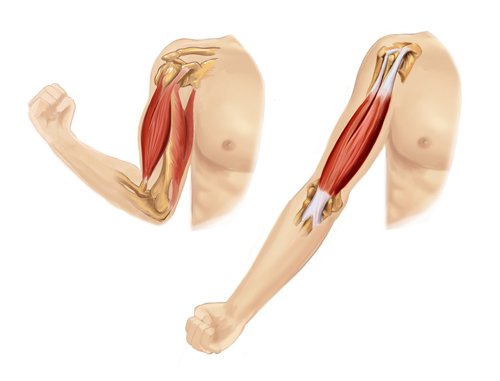
Shivering and shakes are produced in the same way all muscle activity is: by contraction. In fact, all muscle activity is based on contraction. That’s all muscle can do — contract. How do limbs extend, then? The key to more complex actions is that all motion is accomplished by opposing pairs of muscles. Biceps contract to pull your arms up. Triceps, on the other hand, contract to pull your arms down. To clarify our earlier statement, then: all muscle activity is based on opposing contraction. This contraction is initiated by electrical activity. Muscles are highly excitable conductors and respond to electrical stimulation by contracting. In most situations, that electrical stimulation is provided by electrochemical activity in your body. But as anyone who has stuck their finger in a light socket knows, it can also be initiated by outside electrical stimulation.
Types of Muscle
So far, we’ve talked about muscles generically and about the things all muscle tissue shares in common. But the fact is that the body actually contains several different types of muscle that despite any outward similarities are distinctly different in their biomechanics and their uses in the body. The three primary types of muscle are:
- Smooth muscle
- Cardiac muscle
- Skeletal muscle
Smooth muscle is a type of non-striated (un-striped) muscle that is found within the tunica media layer of arteries and veins, the bladder, uterus, male and female reproductive tracts, gastrointestinal tract, respiratory tract, and the ciliary muscle and iris muscles of the eye. The glomeruli of the kidneys also contain a smooth muscle-like cell called the mesangial cell. Smooth muscle is fundamentally different from skeletal muscle and cardiac muscle in terms of structure, function, excitation, excitation-coupling, and its mechanism of contraction.
Cardiac muscle, as its name implies, is found in the walls of the heart, where its contractions propel blood both into the heart and then out through the arteries of the circulatory system. It is similar to skeletal muscle in that it is striated. But unlike skeletal muscle, its contractions are primarily involuntary.
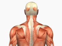
Skeletal muscle is a type of striated muscle that is usually attached to the skeleton by tendons. Skeletal muscles are used to hold posture and create movement, by applying force to bones and joints; via contraction. Unlike smooth and cardiac muscle, they generally contract voluntarily (via somatic nerve stimulation), although they can contract involuntarily through reflexes.
Let’s now look at each of these muscle types in a little more detail — devoting most of our attention to skeletal muscles.
Smooth Muscle
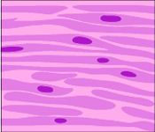 Smooth muscle makes up the walls of hollow organs, hair follicles, and blood vessels. It mostly regulates the size of intestinal muscles and glands and plays the primary role in the contractions of the intestinal tract known as peristalsis. Fundamentally, all muscle tissue is built and powered in the same way, with the difference being that smooth muscle is microscopically smooth, not striated. Instead of being grouped in parallel, smooth muscle cells are assembled in irregular bundles of interwoven clusters. It is called smooth because it doesn’t have the striations found in skeletal muscle. Surprisingly, considering its assembly from irregular bundles, the final construction of the muscle itself tends to be long and slender. It may be innervated (activated) by one nerve or multiple nerves, depending on function. And it is involuntary.
Cardiac Muscle
Cardiac muscle makes up the walls of the heart. It is microscopically striated, like skeletal muscle, but its striations join together in branching bundles that allow coordinated action. The whole unit contracts together VS the selective contraction found in skeletal muscle. And it is involuntary and auto-rhythmic–although, as yogis have demonstrated, even involuntary muscles such as the heart can be made responsive to voluntary control.
Skeletal Muscle
Although skeletal muscle is similar to cardiac muscle and at first glance looks like heart muscle, it has different characteristics…and uses. It is found attached to bone, skin, fascia, and other muscles. Also, any visual similarities to cardiac muscle when viewed with the naked eye disappear when viewed under a microscope. One of the key defining characteristics of skeletal muscle VS cardiac muscle or even the smooth muscle of the intestinal tract is that skeletal muscle is voluntary. That is to say contractions of the skeletal muscle happen when we choose to make them happen — such as when we lift our arms. Cardiac and smooth muscle, on the other hand as we discussed previously, are primarily involuntary. For most people, their hearts tend to beat–or not–no matter what they think about it.
Skeletal muscle looks like it is made up of a series of stripes, which is why it is also sometimes called striated muscle. It has this appearance because it is comprised of a series of long bundled threads of muscle known as myofibrils. It is this bundling together of several groups of fibers in parallel that gives skeletal muscle its striated appearance. Incidentally, myo is Greek for “muscle.”
A single muscle cell is a compound structure composed of several bundles of myofibrils that contain myofilaments. The myofibrils have distinct, repeating microanatomical units, known as sarcomeres, which represent the basic contractile units of the myocyte or muscle cell. (The word sarcomere comes from the Greek: sarco for “flesh” and mere for “part” — thus “flesh part.”)
The appearance of striation is further reinforced by the fact that all of these individual muscle cells and fibers are grouped into parallel bundles known as fascicles, which are wrapped up in a smooth, slippery covering of connective tissue, known as the perimysium. Those bundles are then grouped together and wrapped in a smooth envelope of connective tissue known as the epimysium to form what we know as a single muscle — such as a tricep. From smallest to largest, the parts of a muscle are as follows:
- Sarcomere (the basic contracting unit of the myofilament).
- Myofilament.
- Myofibril.
- Myocyte (a single muscle cell), also referred to as a “muscle fiber.”
- Fascicle (a bundle of 10-100 muscle fibers).
- Perimysium (the layer of connective tissue that surrounds a fascicle).
- A single muscle — such as a bicep.
- Epimysium: The outermost layer of fascia, consisting largely of collagen, surrounding a whole muscle and keeping it distinct from–and allowing it to slide over–adjacent muscle groups.
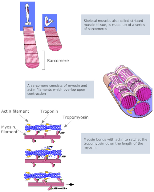
Muscle contraction happens at the level of the sarcomere. The sarcomere is comprised of two protein filaments — the thick filament is made of the protein myosin and the thin filament made of the protein actin. (Incidentally, that’s why protein consumption is important in muscle development. Protein is the fundamental building block in muscle.) When triggered by nerve impulses and powered by ATP, the thick myosin filaments contract. This contraction is transmitted to the thin actin filaments by a series of microscopic spurs in both the thick and thin filaments that ratchet into each other. The thin filaments then transmit the force generated by myosin to the ends of sarcomeres where they are attached and then all along the length of the myofilaments to contract the entire muscle fiber. This is the fundamental element of muscle contraction. Just multiply this out over countless sarcomeres, repeated through vast numbers of fibrils, and numerous muscle fibers, and you have muscle movement. (In a little bit, we’ll talk about how this contraction is powered.)
Motor Unit
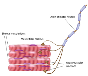 Specific nerves stimulate a specific group of fibers (or muscle cells) referred to as a motor unit. Essentially, a motor unit is composed of a motor neuron and the muscle fibers (cells) it activates. The innervation (stimulation) by a motor neuron may activate as few as 10 muscle fibers to as many as 2,000. The size of that group is determined by the needs of the muscle group in question. The groups are very small in the muscles of the eyes and fingers, for example. This allows for very fine control of those muscles. Groupings in the muscles of the back and thighs, on the other hand, are much larger since there is little need for fine control in those muscles. The signal to activate travels down from the brain, through the spinal cord, and then out to the group of fibers in question. The activating neuron connects to the muscle bundle at what is known as the neuromuscular junction (NMJ). Because most neuromuscular junctions are located in the middle of the muscle fiber, the wave spreads from the middle outward toward the end of the fiber, allowing the muscle to make a smooth contraction. The nerve cell body is located in the spinal cord, with the filament like axon traveling out from the spinal cord to the muscle fibers it controls. The brain can selectively fire muscle fibers as needed by choosing the motor neuron units it wants to fire. All fibers in a motor unit fire if a signal is transmitted through the motor neuron to that unit. Note: the axons are surrounded by a fatty, insulating coating known as the myelin sheath that facilitates the transmission of nerve impulses along the axon and that prevents any leakage of that signal or extraneous signals from disrupting the transmission. Keeping the myelin sheath intact is essential for proper muscle function.
The NMJ actually consists of a microscopic empty space known as a synaptic gap. The electrical impulse of the nerve cannot cross this gap on its own. In place of direct electrical stimulation of the muscle by the nerves, the impulse is carried across the gap by a chemical intermediary: the neurotransmitter. The primary neurotransmitter used in the stimulation of muscle motor units is acetylcholine (Ach), which is produced in synaptic vesicles located in the bulb at the end of the axon. The motor end plate is the region on the muscle side of the synaptic gap in proximity to the axon terminal bulb. The motor end plate contains the Ach receptors, which when touched by the Ach molecule, fire the muscle to contract. Sufficient supplies of acetylcholine are necessary in the synaptic gap for proper muscle function. Ach is carried across the gap by the sodium pump mechanism that we explored in detail when examining the intestinal tract. Thus, maintaining proper hydration and electrolyte balance is essential for proper muscle function.
Acetylcholinesterase is the enzyme that breaks down the Ach and ends the contraction stimulus so the muscle doesn’t spasm. After the process is stopped, there is a refractory period–a period of recovery–in which the muscle is pumping out the sodium, reestablishing the sodium/potassium balance, and repolarizing the charges on either side of the gap–during which no further stimulation is allowed before the muscle fiber can fire again. Fortunately, we have millions of fibers and they’re not all firing together so that while some fibers are recovering, others are ready to go.
Powering Muscles: the ADP-ATP Energy Cycle
Energy is stored in the muscle cells in the form of adenosine triphosphate (ATP). The energy is released when the last phosphate group is split off, converting the ATP into adenosine diphosphate (ADP). The released energy is used to power muscles and do work. This ATP-ADP energy cycle is what powers the ratcheting action in the myosin bands in each individual sarcomere. ADP then needs to be converted back to ATP for another round of work. Rebuilding ATP from ADP requires energy to be pumped back into the system, which is primarily provided by glucose through a process known as glycolysis. Glycolysis is up to 15 times more efficient if oxygen is present.4 Rich PR. “The molecular machinery of Keilin’s respiratory chain.” Biochem Soc Trans. 2003 Dec;31(Pt 6):1095-105. http://www.ncbi.nlm.nih.gov/pubmed/14641005 Anaerobic glycolysis (without oxygen) will produce 2 ATP molecules for every molecule of glucose. But aerobic glycolysis produces 29-30 molecules of ATP for every molecule of glucose. This means that maintaining sufficient oxygenation of muscle tissue is essential for maximum muscle efficiency.
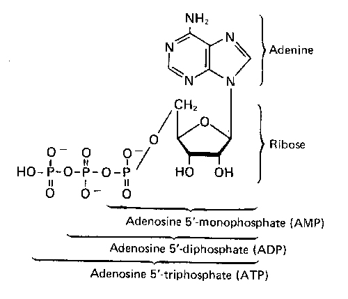
Note: ATP is made up of an adenosine molecule with three attached phosphate groups. Adenosine itself is comprised of a molecule of adenine (one of the key components of your DNA) attached to a ribose sugar. This makes having a sufficient supply of ribose available important for maximum muscle efficiency.
Conclusion
Let’s conclude by detailing those supplements you might want to consider to improve both the capacity of your body to build muscle and its ability to support and maximize the effectiveness of those muscles it already has.
To build muscle tissue:
- HGH secretagogues are precursors necessary for the production of human growth hormone in the body. Typically, such formulas contain ingredients such as glutamine, tyrosine, GABA, arginine, and lysine. HGH promotes the growth of lean muscle in the body, and levels of HGH drop as you age. You don’t want HGH injections for a number of reasons, but a secretagogue supplement will help increase HGH production — as will exercise.
- Protein. Muscle is built from protein, so obviously you need protein to build muscle. But you don’t need massive amounts unless you’re doing extreme levels of exercise, and it doesn’t have to be meat or dairy protein.
- Testosterone unbinders. Testosterone is the hormone that tells your body to use calories to build muscle and not store them as fat. On the other hand, you don’t really want to take additional testosterone, since higher levels of testosterone are associated with a shorter life expectancy in some studies.5 Kyung-Jin Min, Cheol-Koo Lee, Han-Nam Park. “The lifespan of Korean eunuchs.” 25 September 2012. Current Biology 22(18) pp. R792 – R793. http://download.cell.com/current-biology/pdf/PIIS0960982212007129.pdf?intermediate=true Then again, other studies show that low levels of testosterone shorten lifespan.6 Robin Haring, Henry Völzke, Antje Steveling, et al. “Low serum testosterone levels are associated with increased risk of mortality in a population-based cohort of men aged 20–79.” Eur Heart J Feb 17 2010. http://eurheartj.oxfordjournals.org/content/early/2010/02/17/eurheartj.ehq009.full.pdf+html (Let’s hear it for contradictory studies.) Instead, what you want to do is free up the testosterone you already have so that it works more effectively for you. That gives you all of the reward without the risk.
- Carnosine based formulas to protect that muscle from the destructive effects of glycation. Supplementation with carnosine has the additional benefit of dramtically speeding up recovery time after intense workouts.
- Arginine works by filling your muscles with water and nutrients. When taken consistently, it helps your body maintain lean muscle mass and promotes the release of growth hormones.
To allow contiguous muscle groups to easily slide over each other–a healthy epimysium:
- Sufficient hydration. When determining how much water you need, the medical community rarely looks deeply enough and therefore comes to the wrong conclusions. Early stages of dehydration show up in a “sticky” epimysium, which means that muscles no longer glide smoothly over each other.
- Collagen. The epimysium is made largely of collagen. Supplementation with oral collagen supplements helps keep the epimysium intact.
- ASU. Avocado soy unsaponifiables stimulate the production of collagen in the body.
- Systemic proteolytic enzymes help break down any protein adhesions that may form and cause muscles to “stick” together.
- Deep muscle bodywork such as BioSync helps keep the muscle planes from sticking together — and helps separate them if they’re already stuck.
- Yoga. The total body stretches offered in yoga work every muscle plane against every other plane–helping to keep muscle planes moving freely and prevent any adhesions from forming between planes.
To power the nerve impulses that drive muscle activity:
- Acetylcholine. DMAE is a precursor for the production of acetylcholine in the body.
- The myelin sheath is comprised of 70 percent fats and cholesterol and 30 percent protein. It is easily damaged by:
- An accumulation of toxic heavy metals. Detox heavy metals regularly.
- Inflammation in surrounding tissue, which can damage the myelin sheath. Reduce systemic inflammation using proteolytic enzymes
- A deficiency of methylation nutrients compromises the ability of the myelin sheath to repair itself.7 Sangduk Kim1, In Kyoung Lim, Gil-Hong Park, Woon Ki Paik. “Biological methylation of myelin basic protein: Enzymology and biological significance.” The International Journal of Biochemistry & Cell Biology Volume 29, Issue 5, May 1997, Pages 743–751. http://www.sciencedirect.com/science/article/pii/S1357272597000095 Supplementation with any and all of the methylation nutrients such as folic acid, B-12, TMG (tri-methyl-glycine), and SAMe can be helpful.8 Bianchi R, Calzi F, Savaresi S, Sciarretta-Birolo R, Bellasio R, Tsankova V, Tacconi MT. “Biochemical analysis of myelin lipids and proteins in a model of methyl donor pathway deficit: effect of S-adenosylmethionine.” Exp Neurol. 1999 Sep;159(1):258-66. http://www.ncbi.nlm.nih.gov/pubmed/10486194
- Vitamin D protects against demyelination.9 Wergeland S, Torkildsen Ø, Myhr KM, Aksnes L, Mørk SJ, Bø L. “Dietary vitamin D3 supplements reduce demyelination in the cuprizone model.” PLoS One. 2011;6(10). http://www.ncbi.nlm.nih.gov/pubmed/22028844
- Omega-3 fatty acids
Power the ATP-ADP energy process
- CoQ10 and CoQ1 (NADH) are the key members of the electron transfer chain in mitochondria (the primary engines of almost all bioenergy production). The passage of electrons along the electron transport chain is coupled to the formation of ATP. Coenzyme Q-10 needs NADH to be effective.
- Your body generates ATP from D-Ribose, which is normally made from glucose. However if the cell is lacking in energy, then the cell converts the glucose to lactic acid instead of D-Ribose. D-ribose as a nutritional supplement is useful because it is immediately available for the generation of new ATP. Also, sufficient supplies of D-ribose minimize the production of lactic acid.
- Nicotinamide (vitamin B3) plays an important role in the synthesis of components necessary for the production of ATP.
- PQQ (Pyrroloquinoline quinone) is taken as a dietary supplement to support mitochondrial health and cellular energy production and to protect the body from oxidative stress. Even better, it can stimulate production of new mitochondria — where ATP is produced.10 Stites T, Storms D, Bauerly K, et al. “Pyrroloquinoline quinone modulates mitochondrial quantity and function in mice.” J Nutr. 2006 Feb;136(2):390-6. http://jn.nutrition.org/content/136/2/390.full.pdf+html
- Creatine is found naturally in vertebrates and helps to supply energy to all cells in the body.
- Proteolytic enzymes are beneficial here too. Since they improve the blood’s ability to take up oxygen and carry out carbon dioxide, they increase oxygen levels in cells — thus dramatically improving the production of ATP through glycolysis.
That’s it for now. In our next newsletter, we’ll explore the anatomy of the muscular system and how different types of exercise promote the growth of different types of skeletal muscle.
References
| ↑1 | “Hummingbird Heart Rate.” How to Enjoy Hummingbirds. (Accessed 9 Jan 2013.) http://howtoenjoyhummingbirds.com/hummingbird_heart_rate.htm |
|---|---|
| ↑2 | “Shrew.” New World Encyclopedia. (Accessed 9 Jan 2013.) http://www.newworldencyclopedia.org/entry/Shrew |
| ↑3 | Francis G. Benedict and Robert C. Lee. “The Heart Rate of the Elephant.” Proceedings of the American Philosophical Society. Vol. 76, No. 3 (1936), pp. 335-341. http://www.jstor.org/discover/10.2307/984548?uid=2129&uid=2&uid=70&uid=4&sid=21101515429843 |
| ↑4 | Rich PR. “The molecular machinery of Keilin’s respiratory chain.” Biochem Soc Trans. 2003 Dec;31(Pt 6):1095-105. http://www.ncbi.nlm.nih.gov/pubmed/14641005 |
| ↑5 | Kyung-Jin Min, Cheol-Koo Lee, Han-Nam Park. “The lifespan of Korean eunuchs.” 25 September 2012. Current Biology 22(18) pp. R792 – R793. http://download.cell.com/current-biology/pdf/PIIS0960982212007129.pdf?intermediate=true |
| ↑6 | Robin Haring, Henry Völzke, Antje Steveling, et al. “Low serum testosterone levels are associated with increased risk of mortality in a population-based cohort of men aged 20–79.” Eur Heart J Feb 17 2010. http://eurheartj.oxfordjournals.org/content/early/2010/02/17/eurheartj.ehq009.full.pdf+html |
| ↑7 | Sangduk Kim1, In Kyoung Lim, Gil-Hong Park, Woon Ki Paik. “Biological methylation of myelin basic protein: Enzymology and biological significance.” The International Journal of Biochemistry & Cell Biology Volume 29, Issue 5, May 1997, Pages 743–751. http://www.sciencedirect.com/science/article/pii/S1357272597000095 |
| ↑8 | Bianchi R, Calzi F, Savaresi S, Sciarretta-Birolo R, Bellasio R, Tsankova V, Tacconi MT. “Biochemical analysis of myelin lipids and proteins in a model of methyl donor pathway deficit: effect of S-adenosylmethionine.” Exp Neurol. 1999 Sep;159(1):258-66. http://www.ncbi.nlm.nih.gov/pubmed/10486194 |
| ↑9 | Wergeland S, Torkildsen Ø, Myhr KM, Aksnes L, Mørk SJ, Bø L. “Dietary vitamin D3 supplements reduce demyelination in the cuprizone model.” PLoS One. 2011;6(10). http://www.ncbi.nlm.nih.gov/pubmed/22028844 |
| ↑10 | Stites T, Storms D, Bauerly K, et al. “Pyrroloquinoline quinone modulates mitochondrial quantity and function in mice.” J Nutr. 2006 Feb;136(2):390-6. http://jn.nutrition.org/content/136/2/390.full.pdf+html |








Sir:
Sir:
I have “worked out” in the gym for years, but I had stopped for a year or so, then I tried to get back in the gym and “work out,” but I got very dizzy and almost passd out. What do you think is the reason for that happening? Some say it’s because I hadn’t “worked out” for a long while and “shocked” my nervous system; some say it’s because I didn’t eat enough before I “worked out,” which I didn’t. I read that many people have the same problem and they have been checked for their health status and it’s ok, same here. What’s your professional opinion, which I highly respect? Thanks in advance.
Dr. Robert Bolmarcich
Moderate exercisers lose 100%
Moderate exercisers lose 100% of their aerobic capacity in as little as two months of inactivity. Conditioned athletes still lose 50% of their capacity in 3 months. Bottom line: after a year away from the gym, your aerobic capacity would be the same as a couch potato’s. Your brain may remember being fit; your body just doesn’t agree. It will take some time to regain that capacity. Don’t rush it.
Very thorough, thank-you for
Very thorough, thank-you for your work, it is a wonderful resource!
Jon, I have a friend with MS.
Jon, I have a friend with MS. How can I best help her? Whàt products do you have which will help her? I used nto be in Healing America and have met you and Kristen long ago! Thank you!
Sandy, we are sure you are
Sandy, we are sure you are aware that for obvious legal reasons, we cannot diagnose or prescribe for specific medical conditions—merely provide information. With that in mind, you might find the following link useful. https://jonbarron.org/article/ms-and-baseline-health-%C2%AE-program